Pars Fractures of the Spine
Innovative treatments for 3D patching & biologic repair of pars defects

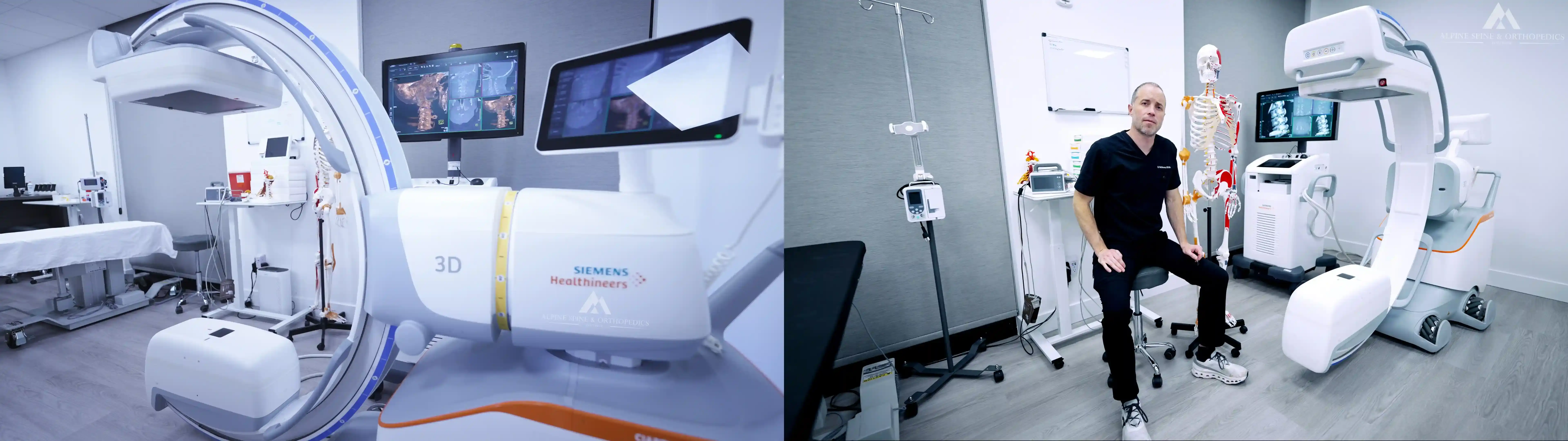
We utilize cutting-edge tissue-engineering approaches to treating pars fractures with “stem cell patching” and "bone marrow grafting" procedures that involve grafting of bone marrow and concentrated stem cell patching with a sticky protein scaffolding matrix precisely placed directly into fractures under high-resolution 3D image-guidance. This form of biological fixation recapitulates the natural way of achieving fracture repair without all the irreversible consequences of hardware implantation. The stem cells used are mesenchymal stem cells (MSCs) and MUSE cells concentrated and grafted from the patient’s own bone marrow, which are already primed for orthopedic tissue repair and which avoid any immunologic rejection response for optimal integration and remodeling (as opposed to foreign cells which express immunologic recognition markers as they integrate and differentiate causing rejection). The stem cells are the same cells that are necessary for all bone fracture repair, just in more concentrated form and with a scaffolding matrix that helps them integrate and remodel injuries over time. We do this procedure under IV sedation and we use minimally-invasive tools like needles, trocars, cannulas, and/or endoscopes with the most advanced high-resolution 3D robotic image-guidance, so there are no incisions nor sutures and patients can walk out afterwards.
STEM CELL PATCHES:
Bone Marrow Grafting + Concentrated Stem Cells + Scaffolding Matrix + Peptide Signaling Factors
The benefits of our 3D-guided stem cell patching procedure include:
1) It is minimally-invasive and far less painful than other spine surgery options (you can go home the same day and generally feels sore for only a short time after the procedure).
2) It does not implant any hardware and does not disrupt the underlying mechanics of the spine, but instead uses a more natural approach (grafting and patching for biological fixation).
3) Multiple issues can be targeted and treated in the same visit, including spine fractures, disc bulges, nerve impingement, facet arthritis, SI joint instability, ligament instability, and so on.
4) In cases of success, it saves patients from the consequences of much bigger surgeries and from a lifetime of painful surgical revisions.
5) In case of failure to repair the fracture, it does not alter any future plans for spine surgery (and in fact, it still helps repair and reinforce surrounding tissues like disc bulges, facet joint cartilage damage, ligament reinforcement, nerve root protection from the fracture edges with hydrogel padding, and so on, which all still help improve function and avoid surgery, as well as reinforce surrounding tissues to help improve outcomes if surgery ever becomes necessary).
Pars fractures are often missed and can cause significant damage even in phases where they do not cause a lot of pain. These fractures put extra strain on discs and facet joints surrounding the fracture, which accelerate a destabilization process in the spine. Many people push through the pain thinking it is just a muscle strain or a disc bulge, and athletes can often play through the pain for a time, but ultimately these injuries rapidly degenerate and typically become career-ending injuries. Yet we have treated dozens of high-level athletes who achieved provable repair of their pars fractures from our innovative procedures, and these athletes have been able to go fully back into demanding sports with no pain and no recurrence of the fractures. This includes many gymnasts, soccer players, football players, basketball players, baseball players, track and field athletes, swimmers, dancers, professional athletes, Olympic athletes, and many others (see case studies below). Regardless of whether active athletes or not, numerous patients have achieved successful repair and resolution of their pain and improved quality of life, including patients who also already had other complex comorbid conditions like disc pathologies, facet arthritis, SI joint instability, connective tissue diseases, hypermobility syndromes, osteoporosis, developmental anomalies like Bertolotti’s syndrome, and many others (many of which we can address with stem cell patching treatment at the same time).
However, it is always possible that the thin pars bone could fracture again after a successful procedure, especially with a fall or with trauma, so caution is still advised. We generally recommend isometric neutral spine with corset bracing for 2-3 months following the procedure, as well as rest for several weeks with gentle progression into core strength maintenance exercises to help prevent atrophy during the healing process, but each case will be given applicable instructions.
There is no spine procedure that has a 100% success rate, so you must be able to accept that this procedure (or any procedure) may not fully repair or resolve the injuries. All spine surgeries have limitations, weak points, and ways that they can fail which typically lead to the need for further spine surgery down the road, and even though most outcome data is never reported for spine surgeries (or only reported in the short-term post-operatively before complications like adjacent segment disease and hardware failures set in), more comprehensive retrospective studies suggest that nearly half of spine surgeries result in some form of failed back surgery syndrome (FBSS). Even good surgeons can have complications like failed hardware, nerve injuries, scar tissue adhesions, CSF leaks, adjacent segment disease (ASD), straight back syndrome (SBS), proximal junctional kyphosis (PJK), lordotic or kyphotic deformities, and other problems with alignment that develop above and below the hardware. Unless patients have seen their own family members go through the agony of spine surgery, most have never seen the many severe complications, consequences, and outcomes that can occur after spine surgery, and even in cases where surgery goes well, once hardware is placed in the spine, the mechanics change and the clock begins ticking for when more advanced revision will become necessary.
The chances of success with this stem cell patching/grafting procedure are much higher if the pars fracture is caught early and if the fracture gap is small. The chances of success are lower if the fracture gap is large, if the fracture is old, if the bone margins are degenerative, sclerotic, or reabsorbed, and if the surrounding structures are collapsed or destabilized with instability and slippage (spondylolisthesis). Nevertheless, many cases of success have involved complete bilateral pars fractures with significant gaps, and we have seen some cases that surprised us with gaps as large as 8mm able to bridge and repair and some cases of success even in older age with long-standing fractures. But there are also failed cases, so in general, if the gap is larger than 4-5mm, if the disc is slipped, collapsed, and unstable, and if the fracture is old degenerative or sclerotic bone with chronically stretched paraspinal ligaments, or if the nerve is being pinched already (typically between the disc and the fracture), then the chances of success drop. We can still attempt to address each of these issues as best as possible without hardware, but it is important to understand the many variables involved and how they relate to each patient’s specific symptoms before making a decision.
Even if the fracture itself is unable to form new bone unionization, there can still be benefits from connective tissue formation and bridging of the fracture gap with a fibrous union by the stem cell patch, because this still stabilizes the spine to some degree to protect against slipping and to reinforce and strengthen surrounding discs and facet joints. In other words, success is not always black and white, and we have had many patients achieve significant improvements in pain and function even when the bone fracture itself has not fully repaired. In some people, the pars fracture itself is the most significant pain generator, but in others, the fracture has led to bigger problems of disc collapse, early facet arthritis, nerve injuries, and so on, so each case is unique and different in many ways, and no single number applies broadly to everyone and no outcome can be guaranteed. Just as with any spine surgery, you cannot predict perfectly who will fall into the ‘success’ vs ‘failure’ group, so we try to analyze all the variables to assess and address each aspect of these injuries in order to achieve maximal repair and the best outcomes possible.
Back pain alone is not a reason to jump to spine surgery, as fusion can often result in even worse back pain, scar tissue, and irreversible consequences above and below the instrumentation. Even bolting the spinal fracture itself, as is done with a direct pars repair or buck screw repair, has many ways that it can fail, especially because it involves placing a small screw through thin bone directly adjacent to nerve roots, and we have seen numerous cases where pars screws have failed to unionize the fracture, failed to improve the pain, caused nerve injury, and/or ultimately loosened in the thin bone over time causing the need for further spine fusion. We even have patients where we grafted stem cell patches around these loosened screws with successful osseointegration in some cases, and even in cases where these screws have achieved unionization, patients have still come to us for patching damaged discs (including disc bulges, fissures, tears, protrusions, and herniations) and for patching facet joints that were damaged by the fractures (especially in cases where the fracture entered into the facet joints). Spinal hardware puts excessive strain and load onto the discs and facet joints around the fracture, so many patients have ended up in worse pain afterwards, and most critically, all rigid fusions and hardware implantation will lead to excess strain and adjacent segment disease above and below the hardware with the need for future revision surgeries over time.
Dr. McMurtrey often operated on some of the most complex spine cases including massive cranium-to-sacrum fusions in patients who had often started down the spine surgery path many years prior due to pars fracture injuries and who had been through numerous revisions surgeries through life, eventually extending the fusion construct all the way up the spine and down into the SI joints. Dr. McMurtrey often thought that if we could go back in time before the first fusion and save even a few of these patients from having to go down this road, then it would be completely life changing for them. He studied biomedical engineering and tissue engineering at the University of Oxford, earning honorary distinction for his work creating 3D tissue structures from patients' own stem cells using guided signaling factors and scaffolding matrix architecture. He is the only doctor who has both surgical training and stem cell research background at top research institutions, and he is currently the only doctor in the world who is using bone marrow grafting, concentrated stem cells, scaffolding matrix, and peptide signaling factors to achieve tissue repair under 3D image-guided techniques. More information on scaffolding matrix, stem cells, and peptides can be found at the links, and each patch composition is customized for each particular tissue and injury.
The following videos give a brief overview of these pars procedures, and several case examples are shown on our Instagram & Facebook pages, including several pars patient case examples posted here: 1 2 3 4 5 6 7 8 9 10 11 12 13 14 15 16 17 18 19 20 21 22 23. Many of our pars fracture patients have also kindly described their experiences and outcomes as found in our reviews.
-----
ASOI © 2024 All Rights Reserved*Disclaimer: The information presented here is for informational use and cites the ongoing cutting-edge research and medical advancements on these relevant topics and advancements. We seek to always provide the highest-quality evidence-based care to our patients customized for their specific conditions, injuries, and diagnoses. The FDA does not regulate the practice of surgery or medicine, but rather regulates medical marketing of devices and drugs. The FDA also does not conduct clinical trials nor do they attempt to discover new treatments. Breakthrough technologies typically require years to decades of research work, which in some cases can optionally be submitted to the FDA if there is sufficient financial backing. Thus the FDA has not yet studied, evaluated, or formally approved many regenerative therapies (nor many other surgical procedures) currently practiced by many of the top physicians and surgeons in the United States and around the world. Some therapies, products, or interventions may still be considered standard practice despite still being "investigational" or "off-label" even with substantial evidence of efficacy, and strangely, many practitioners continue to do some types of procedures even when evidence exists to the contrary (e.g., epidural steroid injections are not FDA-approved and data suggest little benefit but many potential risks of harm, yet approximately 10 million are performed each year in the USA). Common spine surgery techniques also have no specific FDA-approval because the FDA does not regulate surgery nor conduct studies on surgical outcomes. Thus there are many treatments, interventions, and protocols routinely practiced in medicine and surgery which the FDA has not studied nor formally approved yet which have demonstrated overwhelming evidence of efficacy and clinical benefit, while many FDA-approved therapies, standard insurance-covered treatments, and common surgeries can actually have high rates of failure and complication, so patients must always weigh benefits versus risks. Many different applications of regenerative therapies take a newer and more advanced approach to tissue repair and continue to be researched and optimized by our institute and in collaboration with other institutions around the world. In general, treatments may include FDA-approved therapies as well as additional investigational, alternative, or regenerative therapies and interventions, and we always cover potential risks and benefits of these options. The rapid evolution and advancement of surgical and medical interventions demand that physicians continually update their knowledge and practice techniques to adapt to future improvements and advancing technologies for the ultimate benefit of their patients. These statements have not been evaluated by the FDA, and the treatments and products presented here are for educational purposes and are not guaranteed to diagnose, treat, cure, or prevent any specific disease or condition. The information here does not form a doctor-patient relationship nor does it constitute medical advice. All injuries and conditions should be formally evaluated by a knowledgeable medical professional whereby standard treatments and additional therapeutic interventions may be considered with the diagnosis and treatment plan.