Upper Cervical Spine Injuries:
CranioCervical Instability (CCI)
Innovative treatments for complex upper cervical spine injuries & conditions
"Craniocervical Instability" describes a dysfunctional junction between the head and neck. This segment of the spine does not involve discs like the rest of the spine, but instead is composed of ligamentous structures and synovial joints from the base of the skull (C0) to the C2 vertebra. Injuries to these upper cervical segments encompass numerous pathologies and terminologies, including cranio-cervical instability (CCI), atlanto-axial instability (AAI), upper cervical instability, cranial instability, cranio-cervical syndrome, occipito-atlanto-axial hypermobility, and many other associated disorders.
These injuries most commonly occur with trauma and whiplash mechanisms, and symptoms can develop at any time, whether in the acute phase after the injury or many years later as post-traumatic degenerative changes that continue to worsen over time. There is also a significant proportion of CCI patients who also have other associated comorbidities and underlying conditions like subaxial cervical pathology (disc degeneration, facet arthrosis, spondylosis), neurovascular pathologies, connective tissue disorders, hypermobility syndromes, developmental anatomic anomalies, autoimmune conditions, infectious diseases (including Lyme disease, CMV, EBV, etc), and many others. Sometimes conservative treatments like physical therapy, chiropractic therapies, nutritional approaches, and pain management are enough to manage the symptoms. In general, avoid excessive traction on the head and neck and beware steroid injections that can be highly detrimental and worsen these injuries by weakening ligaments, softening cartilage (chondromalacia), inducing bone loss (osteoporosis), etc. When conservative treatments fail, more aggressive therapeutic interventions may become necessary.
STEM CELL PATCHES:
Concentrated Stem Cells + Scaffolding Matrix + Peptide Signaling Factors
We utilize cutting-edge tissue-engineering approaches to treating craniocervical disorders and instabilities with “stem cell patching” procedures that involve concentrated stem cell patching with a sticky protein scaffolding matrix precisely placed directly onto cranio-cervical joint cartilage and capsules (C0-1 & C1-2) and cranio-cervical ligaments (including critical alar and transverse ligament patching via anterior trans-oral approaches to the cranio-vertebral junction) under high-resolution 3D image-guidance. This form of biological patching recapitulates the natural way of achieving tissue repair without all the irreversible consequences of hardware implantation. The stem cells used are mesenchymal stem cells (MSCs) and MUSE cells concentrated and grafted from the patient’s own bone marrow, which are already primed for orthopedic tissue repair and which avoid any immunologic rejection response for optimal integration and remodeling (as opposed to foreign cells which express immunologic recognition markers as they integrate and differentiate causing rejection). The stem cells are combined with a scaffolding matrix that reinforces the ligaments and anchors the stem cells directly to the targeted injury sites so they do not leak back out or undergo anoikis. The protein scaffolding matrix acts as a form of biologic glue that also optimizes integration with the injured tissues, can be infused with additional peptide signaling factors that diffuse over time, and directly activates platelets with fibrin-platelet integrin receptors to stimulate release of growth factors. We do this procedure under IV anesthesia and we use minimally-invasive tools like needles, trocars, cannulas, tubular retractors, and endoscopes with the most advanced high-resolution 3D robotic image-guidance, so there are no incisions nor sutures and patients can walk out afterwards.
These procedures are complex and must be customized for each patient’s specific and unique set of injuries, and we typically require at minimum a cervical MRI and a digital motion x-ray (DMX) of the upper cervical segments before we can evaluate your case for these procedural interventions. The doctor reviews this imaging in great detail and correlates your symptoms to the pathologies seen in order to best treatment plan your case. We also suggest first trying all conservative measures to strengthen core muscle support and regain proper postural correction before proceeding with more invasive interventions.
We have numerous patients who have achieved dramatic improvements with resolution of their symptoms after just a single treatment. Nevertheless, each case is unique and involves numerous variables, so just as in spine surgery, it cannot always be predicted who will do great versus who may need additional treatments. The repair process takes time and occurs in phases rather than a straight linear path, and most patients have achieved significant benefits by 3 months, with benefits still lasting multiple years now in most of our early cases. However, in more severe cases, more than one treatment may be needed, or surgical fusion may be necessary as a last resort.
As CCI progresses, the problem becomes not just the worsening instability itself, but the consequences on adjacent tissues as a result of the instability, including accelerated wear of the cartilage in the C0-1 & C1-2 joints, development of early arthritis and bone spurs, excessive strain on paraspinal ligaments, excessive stretching of nerves (causing injury to nerve roots, peripheral nerves, cranial nerves, and autonomic nerves), strain on the underlying disc and facets, neurovascular injury particularly to vertebral arteries passing through these segments (such as vertebral dissection), and ultimately involvement of critical spinal cord and brain stem structures. Fortunately we have treated many types of severe injuries like complex odontoid (dens) fractures, severe atlantoaxial arthritis, traumatic subluxation with vertebral artery dissection, and basilar invagination cases that have had great success, but those patients’ outcomes cannot be broadly applied or predictive for every unique case.
Some patients have said they felt better very quickly after treatment with even further sustained benefits over time, which is likely related to the hydraulic hydrolocking hydrogel effect of the scaffolding matrix on the instability and cushioning the joint cartilage and capsules, similar to how a flat tire becomes more supported, more stable, and better functioning when reinflated with green goo to seal a leak. In this case, the ‘goo’ is the biologic glue of sticky protein scaffolding matrix, and it not only provides mechanical benefits but also promotes tissue integration and provides a matrix to rebuild on. In the lab, stem cells thrive on protein scaffolding matrices (for example, matrigel is often used as a matrix in 3D stem cell cultures in the lab, which has dramatically different effects versus culturing cells in PRP-like solutions where stem cells tend to undergo a programmed cell death called anoikis).
Dr. McMurtrey studied biomedical engineering and tissue engineering at the University of Oxford, earning honorary distinction for his work creating 3D tissue structures from patients' own stem cells using guided signaling factors and scaffolding matrix architecture. While most doctors presently doing stem cell treatments are just rehab doctors and pain management doctors with no surgical training and no formal stem cell research background, Dr. McMurtrey is the only doctor who has both surgical training and stem cell research background at top research institutions, including University of Virginia where the Journal of Neurosurgery is published and which has trained numerous chairmen of neurosurgery, as well as University of Oxford which is consistently ranked as the world’s #1 research university, with top ranking of the biomedical engineering department and where some of the most significant stem cell research discoveries have been made. Dr. McMurtrey is currently the only doctor in the world who is using concentrated stem cells, scaffolding matrix, and peptide signaling factors to achieve tissue repair under 3D image-guided techniques. More information on scaffolding matrix, stem cells, and peptides can be found at the links, and each patch composition is customized for each particular tissue and injury.
Trans-oral and trans-nasal approaches to the cervical spine have been done for well over 100 years, including to the pituitary by Harvey Cushing in the 1900s then craniovertebral pathologies as early as 1917 for atlantoaxial subluxation as well as resection of tumors, tuberculosis, and even a bullet. Then in the 1980s Dr. Arnold H. Menezes significantly advanced these approaches and techniques to dramatically improve both safety and efficacy to make this approach the mainstay for these unique pathologies. Dr. McMurtrey trained with some of the leading experts in craniocervical surgery for atlantoaxial instabilities, basilar invagination, and craniocervical subluxation, including operating with Dr. Arnold Menezes, Dr. John Jane Sr, and Dr. Edward Oldfield on numerous craniocervical fusions and endoscopic trans-oral and trans-nasal cases in both pediatric and adult cases. Craniocervical fusion carries many significant risks with permanent irreversible consequences and major potential complications. Even if the fusion surgery goes well, many patients have still said they felt worse, and the hardware will always lead to excess strain on the adjacent segments often leading to revision and extension of fusion, and fusion fails to address additional adjacent tissue injuries that may also contribute to symptoms, so we try to be as comprehensive as possible in identifying the underlying pathologies and treatment targets so that we can do everything possible to help avoid the fusion path.
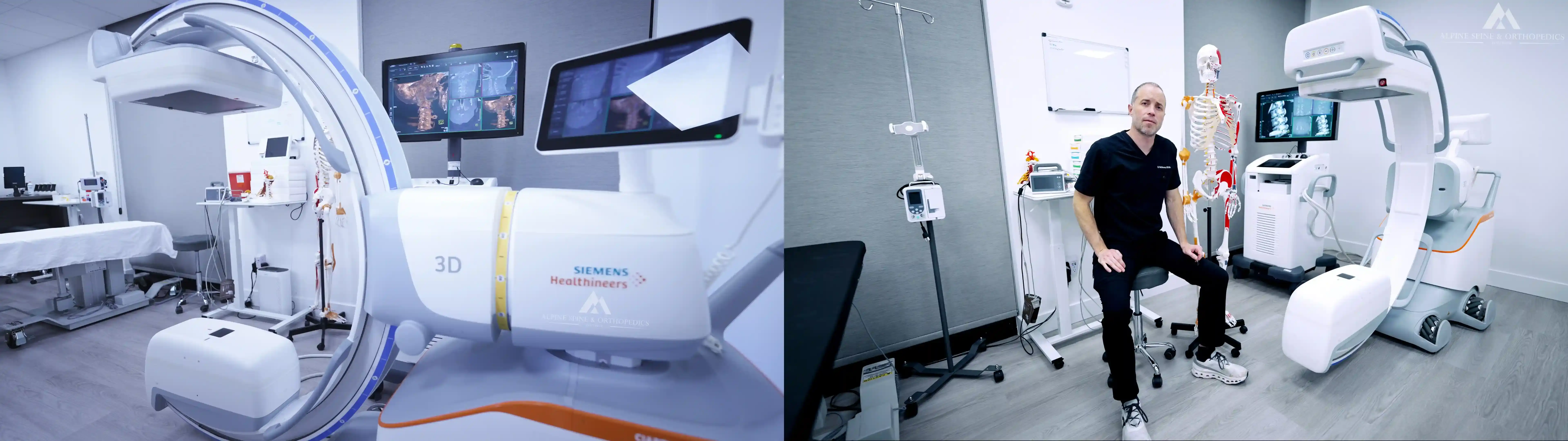
Our high-resolution 3D image-guidance enables never-before-seen detail and resolution of the anatomy and placement of the stem cell patches. No other place in the world is capable of this level of detail and accuracy. Other providers have traditionally used basic X-ray guidance with contrast dye injections, which unfortunately uses dye to fill up the small space available. X-ray images also compress overlapping bone and spaces into a single vague 2D image where it cannot be determined precisely where the needle is or whether it has accidentally penetrated ligaments or CSF space. It is not surprising that actual procedural images of these anterior cervical injections are not typically shown by any other providers, and the very few that have surfaced have been extremely low quality. We are the only clinic that has shown immaculate examples of perfectly accurate targeting using high-resolution 3D imaging in multi-plane slices and perfect placement of patches filling the space around the ligaments and filling in cartilage defects of the C1-2 and C0-1 joints without any displacement by contrast dye (see numerous examples below). Our advanced imaging has often revealed additional findings as well, including bone spurs, ossicles, nerve impingements, vascular impingements, arch defects, dysplasia, tropisms, and other anatomic anomalies.
The following videos give a brief overview of these CCI procedures, and several case examples are shown on our Instagram & Facebook pages, including several CCI patient case examples posted here: 1 2 3 4 5 6 7 8 9 10 11 12. Many of our upper cervical CCI patients have also kindly described their experiences and outcomes as found in our reviews.

-----
ASOI © 2024 All Rights Reserved*Disclaimer: The information presented here is for informational use and cites the ongoing cutting-edge research and medical advancements on these relevant topics and advancements. We seek to always provide the highest-quality evidence-based care to our patients customized for their specific conditions, injuries, and diagnoses. The FDA does not regulate the practice of surgery or medicine, but rather regulates medical marketing of devices and drugs. The FDA also does not conduct clinical trials nor do they attempt to discover new treatments. Breakthrough technologies typically require years to decades of research work, which in some cases can optionally be submitted to the FDA if there is sufficient financial backing. Thus the FDA has not yet studied, evaluated, or formally approved many regenerative therapies (nor many other surgical procedures) currently practiced by many of the top physicians and surgeons in the United States and around the world. Some therapies, products, or interventions may still be considered standard practice despite still being "investigational" or "off-label" even with substantial evidence of efficacy, and strangely, many practitioners continue to do some types of procedures even when evidence exists to the contrary (e.g., epidural steroid injections are not FDA-approved and data suggest little benefit but many potential risks of harm, yet approximately 10 million are performed each year in the USA). Common spine surgery techniques also have no specific FDA-approval because the FDA does not regulate surgery nor conduct studies on surgical outcomes. Thus there are many treatments, interventions, and protocols routinely practiced in medicine and surgery which the FDA has not studied nor formally approved yet which have demonstrated overwhelming evidence of efficacy and clinical benefit, while many FDA-approved therapies, standard insurance-covered treatments, and common surgeries can actually have high rates of failure and complication, so patients must always weigh benefits versus risks. Many different applications of regenerative therapies take a newer and more advanced approach to tissue repair and continue to be researched and optimized by our institute and in collaboration with other institutions around the world. In general, treatments may include FDA-approved therapies as well as additional investigational, alternative, or regenerative therapies and interventions, and we always cover potential risks and benefits of these options. The rapid evolution and advancement of surgical and medical interventions demand that physicians continually update their knowledge and practice techniques to adapt to future improvements and advancing technologies for the ultimate benefit of their patients. These statements have not been evaluated by the FDA, and the treatments and products presented here are for educational purposes and are not guaranteed to diagnose, treat, cure, or prevent any specific disease or condition. The information here does not form a doctor-patient relationship nor does it constitute medical advice. All injuries and conditions should be formally evaluated by a knowledgeable medical professional whereby standard treatments and additional therapeutic interventions may be considered with the diagnosis and treatment plan.